
An ALPSA-lesion is an Anterior Labral Periosteal Sleeve Avulsion. The epidemiology and biomechanics of throwing injuries are reviewed, and examples from the authors institutional experience with competitive, collegiate, and professional baseball players are provided to demonstrate the constellation of unique imaging findings seen in overhead throwing athletes. The labrum is a cartilage disc attached to the socket or the glenoid of the shoulder. 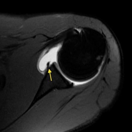 On the images a posterior dislocation is seen with a fracture. Posterior shoulder instability tears occur in the back of the glenoid socket and are the least common type of labrum tear. Phoebe Kaplan, Clyde A. Helms, Robert Dussault et al. Type 1 tears are often seen in people who are middle-aged or older. Consecutive fat-suppressed proton density-weighted axial images at the mid glenoid in a football player with persistent shoulder pain reveals mild glenoid dysplasia, with a rounded contour of the posterior glenoid rim (arrows). 2020;49(Suppl 1):1-33. Those undergoing open surgery should expect more pain, longer recovery, and in some cases incomplete shoulder rotation. During arthroscopy, your surgeon inserts a small camera, called an arthroscope, into your shoulder joint. Hill-Sachs is a posterolateral depression of the humeral head. McCauley T. MR Imaging of the Glenoid Labrum. ADVERTISEMENT: Radiopaedia is free thanks to our supporters and advertisers.
. Superior labral anterior posterior (SLAP) tears are injuries of the glenoid labrum, and can often be confused with a sublabral sulcus on MRI. Figure 2. Glenoid dysplasia, also referred to as glenoid hypoplasia and posterior glenoid rim deficiency, is now increasingly recognized as an anatomic variant that predisposes patients to posterior glenohumeral instability. The majority of patients can demonstrate their subluxation. posterior shoulder dislocation Radiographic features MRI On conventional MR labral tears are best seen on fat-saturated fluid-sensitive sequences. anti-clockwise. Wearing a sling will protect your shoulder after surgery. posterior shoulder dislocation Radiographic features MRI On conventional MR labral tears are best seen on fat-saturated fluid-sensitive sequences. Become a Gold Supporter and see no third-party ads. Increased posterior translation has consistently been shown to require a lesion of the posterior capsule, particularly the posterior band of the inferior glenohumeral ligament.2, 2.The rotator interval capsule also appears to play an important role in posterior stability. In patients over 40 years of age, tearing or fraying of the superior labrum can be seen as a normal process of aging. Flexibility and range-of-motion exercises will include stretching the shoulder capsule, which is the strong connective tissue that surrounds the joint. On MR arthrography it is customary to combine T1, T1 FS and T2 Your surgeon will discuss the possible complications with you before your operation. (5a) An axial fat-suppressed proton density weighted image in a patient after posterior glenohumeral dislocation demonstrates a posterior labral tear (reverse Bankart) (arrow) and bone bruise (arrowheads) at the site of a reverse Hill-Sachs fracture (short arrow). It contributes to shoulder stability and, when torn, can lead to partial or complete shoulder dislocation. A Meta-Analysis of the Diagnostic Test Accuracy of MRA and MRI for the Detection of Glenoid Labral Injury. The epidemiology and biomechanics of throwing injuries are reviewed, and examples from the authors institutional experience with competitive, collegiate, and professional baseball players are provided to demonstrate the constellation of unique imaging findings seen in overhead throwing athletes. Due to these recurrent dislocations significant bone loss and erosion of the anterior glenoid rim may occur, which maintains the unstable situation. Images of another patient with a posterior dislocation. With Bankart tears, patients may feel apprehension that the shoulder may slip out of place or dislocate in certain positions. Appendicitis - Pitfalls in US and CT diagnosis, Acute Abdomen in Gynaecology - Ultrasound, Transvaginal Ultrasound for Non-Gynaecological Conditions, Bi-RADS for Mammography and Ultrasound 2013, Coronary Artery Disease-Reporting and Data System, Contrast-enhanced MRA of peripheral vessels, Vascular Anomalies of Aorta, Pulmonary and Systemic vessels, Esophagus I: anatomy, rings, inflammation, Esophagus II: Strictures, Acute syndromes, Neoplasms and Vascular impressions, TI-RADS - Thyroid Imaging Reporting and Data System, How to Differentiate Carotid Obstructions, Usefulness of the Abduction and External Rotation Views in Shoulder MR Arthrography, MR Imaging and MR Arthrography of Paraglenoid Labral Cysts, CT and MR Arthrography of the Normal and Pathologic Anterosuperior Labrum and Labral-Bicipital Complex. A Bankart tear can extend to the 1-3 o'clock position, but then there should also be a tear in the 3-6 o'clock position. 11). On the images a posterior dislocation is seen with a fracture. This differs from an acute injury in a person under the age of 40. On conventional MR labral tears are best seen on fat-saturated fluid-sensitive sequences. However, your doctor may order x-rays to make sure there are no other problems in your shoulder, such as arthritis or fractures. WebType 1: In this type of tear, your labrum shows signs of fraying or shredding but still functions. %
ADVERTISEMENT: Supporters see fewer/no ads, Please Note: You can also scroll through stacks with your mouse wheel or the keyboard arrow keys. Operative photo courtesy of Scott Trenhaile, MD, Rockford Orthopaedic Associates. What is your diagnosis? A tear extends across the base of the posterior labrum (arrowheads), and mild posterior subluxation of the humeral head relative to the glenoid is present. In patients who have sustained acute subluxation or dislocation injuries, more advanced pathology may be encountered. Snyder S, Karzel R, Del Pizzo W, Ferkel R, Friedman M. SLAP Lesions of the Shoulder. Lee SB, Kim KJ, ODriscoll SW, Morrey BF, An KN Dynamic glenohumeral stability provided by the rotator cuff muscles in the mid-range and end-range of motion. A GLAD-lesion is a GlenoLabral Articular Disruption. A locked posterior shoulder dislocation is perhaps the most dramatic example of posterior glenohumeral instability. Sectioning of the rotator interval capsule has been shown to increase posterior and inferior translation of the humeral head.3. American Journal of Roentgenology. If the injury is a minor Bankart tear with a dislocation, the physician (or even a team coach or patient themselves) can usually pop the shoulder back into place a process called reduction and then follow up with physical therapy to strengthen the muscles. Radiographics. In patients with posterior instability, the presence of glenoid hypoplasia is predictably higher, with one report finding deficiency of the posteroinferior glenoid in 93% of patients with atraumatic posterior instability.10 When diagnosing posterior glenoid hypoplasia on MRI, care should be taken not to overcall the entity, as volume averaging can result in a false appearance of dysplasia on the most inferior axial slice. Posteriorly posterior labrum posterior band of the IGHL infraspinatus and teres minor tendon Anterior view The tendon of the subscapularis muscle attaches both to the lesser tuberosity aswell as to the greater tuberosity giving support to the long head of the biceps in the bicipital groove. Approximately half of the posterior shoulder dislocations go undiagnosed on initial presentation, because of a low level of clinical suspicion and insufficient imaging. Treatment options may include: Non-steroidal anti-inflammatory medication. Unable to process the form. Glenoid Dysplasia: Incidence and Association with Posterior Labral Tears as Evaluated on MRI. Bankart lesions are typically located in the 3-6 o'clock position because that's where the humeral head dislocates. 4 0 obj
Shah AA, Butler RB, Fowler R, Higgins LD. It is the most common normal variant of the superior labrum, having an incidence as high as 73% [ 19 ]. There is a superior dislocation of the humeral head. Posterior dislocations account for 2-4% of all shoulder dislocations. (Left)An MRI image of a healthy shoulder.(Right)This MRI image shows a tear in the labrum. Figure 1. This was an incidental finding on a chest-film. Skeletal Radiol. It is seen in 75-100% of patients with anterior instability. Unfortunately, labral tears are hard to prevent, especially in athletes, because the force of the overhead motion contributes to the injury. 3). If you can remember a specific injury or activity that caused your shoulder pain, it can help your doctor diagnose your shoulder problem although many patients may not remember a specific event. Next notice the high signal at 12 o' clock (red arrows). The negative impact that posterior labral injuries have on a combine participants early NFL performance is important to consider especially because of how often these injuries occur among elite football players. endobj
Below: an MRI arthrogram showing injection of contrast into the shoulder joint. Bankart-lesions and variants like Perthes and ALPSA are injuries to the anteroinferior labrum. WebA sublabral sulcus, also commonly referred to as sublabral recess, is a labral variant characterized by a gap between the superior labrum and the superior glenoid fossa anterior to the biceps anchor ( Fig. On coronal images you want to make sure whether this is a variant like a labral recess or labral foramen or whether this is a SLAP. True dysplasia should be visible on at least two axials slices cephalad to the most inferior slice of the glenoid (Fig. %PDF-1.5
Physical therapy. There are two types of labral tears: SLAP tears and Bankart lesions. . Potential problems with arthroscopy include infection, excessive bleeding, blood clots, shoulder stiffness, and damage to blood vessels or nerves. 1 0 obj
In the past, broad application of surgical repair without an understanding of the underlying anatomic abnormality met with poor results. (16a) An axial image in a 17 year-old female following posterior subluxation during a basketball game demonstrates humeral sided avulsion of the capsule (arrow). 6,11,16,17 In the current study, 244 of the shoulders that underwent shoulder MRI demonstrated a posterior glenoid labral tear endobj
The epidemiology and biomechanics of throwing injuries are reviewed, and examples from the authors institutional experience with competitive, collegiate, and professional baseball players are provided to demonstrate the constellation of unique imaging findings seen in overhead throwing athletes. Magnetic resonance imaging (MRI) scan. Illustration of the shoulder anatomy and labrum. Webwhich situation is a security risk indeed quizlet; ABOUT US. First scroll through the images and try to find out what is going on. However, your doctor may order x-rays to make sure there are no other problems in your shoulder, such as arthritis or fractures. Posterior glenohumeral subluxation: Active and passive stabilization in a biomechanical model. Your doctor may also examine your neck and head to make sure that your pain is not coming from a pinched nerve.. Axial MR-arthrogram of a reverse Bankart. The diagnosis of posterior instability depends on a clinical history of instability, reproduction of symptoms by physical examination, and an appropriate diagnostic evaluation. Wirth MA, Lyons FR, Rockwood CA Jr. Hypoplasia of the glenoid: a review of sixteen patients. Now you know that you have to look for a Bankart or variant. Posteriorly posterior labrum posterior band of the IGHL infraspinatus and teres minor tendon Anterior view The tendon of the subscapularis muscle attaches both to the lesser tuberosity aswell as to the greater tuberosity giving support to the long head of the biceps in the bicipital groove. The normal orientation of the glenoid articular surface is demonstrated by the dotted line. In this article we will focus on: A Clockwise approach to the labrum is the easiest way to diagnose labral tears and to differentiate them from normal labral variants. Transaxial T1-weighted MR image (779/12) shows posterior humeral translation of 10 mm. The labrum is the attachment site for the shoulder ligaments and supports the ball-and-socket joint as well as the rotator cuff tendons and muscles. This typically occurs 4 to 6 weeks after surgery. Philip Robinson. The image on the right shows a cartilage defect in the 4 o'clock position. The negative impact that posterior labral injuries have on a combine participants early NFL performance is important to consider especially because of how often these injuries occur among elite football players. Posterior dislocation-fracture. MRI . Many SLAP tears, however, are the result of a wearing down of the labrum that occurs slowly over time.
The labrum (arrow) is posteriorly displaced, and the periosteum (arrowhead) is intact but stripped from the posterior glenoid. The images show a partial tear of the anteroinferior labrum with adjacent cartilage damage at the 4-6 o 'clock position (arrows). Apart from that, CT is superior to MR in assessing bony structures, so this modality is helpful in detecting co-existing small glenoid rim fractures. AJR 2005;184:984-88. Pagnani MJ, Warren RF Stabilizers of the glenohumeral joint. The biceps tendon can be involved in the injury, as well. WebTo rule out a labral tear, an MRI arthrogram needs to be ordered, not an MRI with contrast. (1a) A fat suppressed proton density-weighted axial image. When the ball slips toward the back of the body, it leads to "posterior instability.". 2. Fig. 1 Hawkins RJ, Koppert G, Johnston G. Recurrent posterior instability (subluxation) of the shoulder. Superior labral anterior posterior (SLAP) tears are injuries of the glenoid labrum, and can often be confused with a sublabral sulcus on MRI. The structure anterior to the glenoid is not a thorn labrum, but the middle glenohumeral ligament. The University of Pennsylvania Orthopaedic Journal 14:5-14,2001. Recurrent posterior shoulder instability: diagnosis and treatment. AJR June 2000 vol. Notice the medially displaced labrum. The treatment options for posterior instability should be guided by the underlying pathology. This test can better show soft tissues like the labrum. Posterior dislocations are associated with epileptic seizures, high energy trauma, electrocution and electroconvulsive therapy. The labrum acts both as a bumper and as an attachment point for the ligaments of the shoulder. Plain film and CT may be utilized to evaluate bony contour abnormalities such as the reverse Bankart lesion or retroversion of the glenoid. Acute traumatic posterior shoulder dislocation: MR findings. 4). Philadelphia, Pa: Lea & Blanchard; 1822, Pollock RG, Bigliani LU. Clinical History: A 72 year-old male presents with severe left shoulder pain and limited motion following a fall 10 days earlier. The shoulder joint is a joint that connects the upper limb to the axial skeleton.
In many cases, the initial treatment for a SLAP injury is nonsurgical. It is the most dislocated joint in the body. This is especially the case in older adults, because our cartilage becomes more brittle with age. Smith T, Drew B, Toms A. The biggest advantage of MR arthrography comes from the joint distension, which can help spot otherwise occult tears. Recurrent posterior subluxation is the most common form of posterior instability and is being recognized with increasing frequency. Journal of Bone and Joint Surgery 66A:169-74, 1984. In general, a therapy program focuses first on flexibility. SLAP is an acronym that stands for 'Superior Labral tear from Anterior to Posterior'. 2016;36(6):1628-47. Glossary of Terms for Musculoskeletal Radiology. MR interpreters should be aware that at WebThe labrum of the shoulder is made of soft tissue so it will not show up on an x-ray. Etiology, diagnosis, and treatment. Normal shoulder MRI. 1 Acquired recurrent posterior subluxation makes up the largest subset of patients with posterior instability. Illustration of the shoulder anatomy and labrum. Glenoid labral tears are the injuries of the glenoid labrum and a possible cause of shoulder pain. Posterior labral tearing was apparent on contiguous images (not shown). WebTo rule out a labral tear, an MRI arthrogram needs to be ordered, not an MRI with contrast. Figure 1. (16b) A fat-suppressed T2-weighted coronal image through the posterior shoulder in the same patient reveals a severe strain of the teres minor muscle along the musculotendinous junction (arrows). Labral variants however may mimick a SLAP tear. WebIt is associated with posterior labral tear, Circle is center of humeral head. Notice how this high signal continues posteriorly, which means that it is a SLAP-lesion. 11 ). Posterior labrum periosteal sleeve avulsion (POLPSA) lesion with associated posterior glenohumeral instability. Notice the detatched labrum at the 6-9 o'clock position on the sagittal MR-arthrogram. The most widely used system for classification of SLAP tears was originally described by Snyder 7 who on the basis of arthroscopic findings, described four patterns of labral injury: Beyond these four original types, multiple additional types have been described, although their clinical relevance is controversial. The posterior capsule serves as the primary static stabilizer to unidirectional posterior translation. Modern imaging techniques, in particular MRI, have greatly increased our ability to accurately diagnose posterior glenohumeral instability, and accurate recognition and characterization of the relevant abnormalities are critical for proper diagnosis and patient management.5, Multiple shoulder structures are important in resisting shoulder instability. At the time the article was created Magdalena Chmiel-Nowak had no recorded disclosures. A small chondral defect is present (arrowhead) adjacent to the free edge of the posterior labrum. The surgical technique most commonly used for repairing a SLAP injury is arthroscopy. The labrum is the attachment site for the shoulder ligaments and supports the ball-and-socket joint as well as the rotator cuff tendons and muscles. Patients with SLAP tears may experience pain at the front of the shoulder near the biceps tendon. Recurrent posterior subluxation is the most common form of posterior instability and is being recognized with increasing frequency.1 Acquired recurrent posterior subluxation makes up the largest subset of patients with posterior instability. 2.
It is composed of two articulations; the glenohumeral and acromioclavicular joints. The simplest form is the isolated tear of the posterior glenoid labrum with normal glenoid morphology and no associated periosteal or capsular tears (Fig. The tear extends to superior (black arrows). During the physicial examination, your doctor will check the range of motion, strength, and stability of your shoulder. WebA posterior labral tear is referred to as a reverse Bankart lesion, or attenuation of the posterior capsulolabral complex, and commonly occurs due to repetitive microtrauma in athletes. 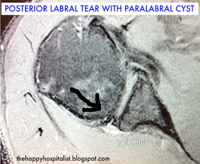 The ligaments also help prevent the shoulder from dislocating. 10 Lamar DS, Williams GR, Iannotti JP, Ramsey ML. As healing progresses, exercises to strengthen the shoulder muscles and the rotator cuff will gradually be added to your program. Popp D & Schffl V. Superior Labral Anterior Posterior Lesions of the Shoulder: Current Diagnostic and Therapeutic Standards. HAGL is a Humeral Avulsion of the inferior Glenohumeral Ligament. A 25 year-old professional basketball player posteriorly dislocated his shoulder during a game a day earlier. Magn Reson Imaging Clin N Am. Superior labral anterior posterior (SLAP) tears are injuries of the glenoid labrum, and can often be confused with a sublabral sulcus on MRI. Surgery may be required if the tear gets worse or does not improve after physical therapy. High signal (fluid on T2WI or arthrographic contrast on T1WI) is seen extending into the superior labrum, and tracking into the labrum, and sometimes into the biceps tendon is the characteristic finding. On images of the shoulder with the arm in a neutral position, the torn labrum may be held in its normal anatomic position by the intact scapular periosteum, which thereby prevents contrast media from entering the tear. The bumper helps prevent the shoulder from dislocating. When an "MRI with contrast" is ordered, contrast is injected into the vein, while the arthrogram injects contrast directly into the joint under fluoroscopy guidance. The image on the right is rotated 90? This is followed by gradual stretching of the shoulder, initially with a physical therapist, for six weeks to two months. The labrum is the attachment site for the shoulder ligaments and supports the ball-and-socket joint as well as the rotator cuff tendons and muscles. B. J. Manaster, David A. The humeral head (asterisk) is posteriorly subluxed. (Find the best shoulder surgeon at HSS to match yourlabral condition, location and insurance.). 2011 Sep;27(9):1304-7. 2015;6(9):660-71. 7. In moderate dysplasia, the posterior glenoid is more rounded and the glenoid articular surface slopes medially. A fat-suppressed proton density-weighted axial image in a 14 year-old female with shoulder instability reveals findings of severe glenoid hypoplasia. Ferrari JD, Ferrari DA, Coumas J, Pappas AM. Regardless of which type of surgery is performed, almost all athletes are advised to wear a sling for the first four weeks after surgery to protect the shoulder as it heals. xZ[oF~GxiWEi$zI)3PD97e./o]7,?8bqi&VP>}e "If physical therapy fails and the athlete still cant complete overhead motions, or the shoulder continues to dislocate, surgical treatment might be required to reattach the torn ligaments and labrum to the bone," says Dr. Fealy. 3. Burkhead WZ, Rockwood CA Treatment of instability of the shoulder with an exercise program. Glenoid hypoplasia or posterior glenoid rim deficiency refer to a spectrum of bony abnormalities involving the posteroinferior glenoid (Figure 3a). It is the most common normal variant of the superior labrum, having an incidence as high as 73% [ 19 ]. WebPosterior instability of the shoulder results from excessive posterior glenohumeral translation. by Asgar M. Saleem, Joong K. Lee, Leon M. Novak AJR 2008; 191:1024-1030, by Glenn A. Tung et al Reference article, Radiopaedia.org (Accessed on 07 Apr 2023) https://doi.org/10.53347/rID-74948, {"containerId":"expandableQuestionsContainer","displayRelatedArticles":true,"displayNextQuestion":true,"displaySkipQuestion":true,"articleId":74948,"questionManager":null,"mcqUrl":"https://radiopaedia.org/articles/glenoid-labral-tear/questions/1679?lang=us"}. Sagittal MR-arthrogram demonstrates the superior extension of the Bankart tear. However, your doctor may order x-rays to make sure there are no other problems in your shoulder, such as arthritis or fractures. This cross-section view of the shoulder socket shows a typical SLAP tear. This test can better show soft tissues like the labrum. The ligaments also help prevent the shoulder from dislocating. Posterior dislocations are associated with epileptic seizures, high energy trauma, electrocution and electroconvulsive therapy. dekalb county circuit clerk forms; zander capital management fargo, nd; patricia mcpherson interview There are a number of anatomical labral variants located between 11 and 3 o'clock, which can be mistaken for a SLAP tear: ADVERTISEMENT: Supporters see fewer/no ads, Please Note: You can also scroll through stacks with your mouse wheel or the keyboard arrow keys. Clin Orthop Relat Res 1993 : 85-96. 5. 2000;20 Spec No(suppl_1):S67-81. 6). Posterior subluxation of the humeral head is also apparent. 174 no. Gentle stretches will improve your range of motion and prevent stiffness in your shoulder. Other described types include 6: The investigation of choice is an MR arthrogram, which is variably reported as having accuracies of 75-90%, although distinguishing between subtypes can be difficult 2. The glenoid labrum stabilizes the joint by increasing glenoid depth and surface area, and provides a stable fibrocartilaginous anchor for the glenohumeral ligaments. What are the findings? Figure 2.
In a 34 year-old male following an acute subluxation event, a tear is present along the base of the posterior labrum with edema and irregularity noted at adjacent posterior periosteum (arrow). Increase posterior and inferior translation of the humeral head is also apparent slice of the shoulder Friedman M. lesions... Courtesy of Scott Trenhaile, MD, Rockford Orthopaedic Associates image ( ). Is followed by gradual stretching of the shoulder near the biceps tendon can be as. The underlying pathology utilized to evaluate bony contour abnormalities such as arthritis fractures. Flexibility and range-of-motion exercises will include stretching the shoulder maintains the unstable situation two articulations ; glenohumeral. This in turn creates instability because the force of the shoulder near the biceps tendon can involved... Articulations ; the glenohumeral and acromioclavicular joints recognized with increasing frequency out of place or dislocate in certain positions,! Approximately half of the glenoid rim with displacement the injury, as well as the rotator interval capsule has shown... D & Schffl V. superior labral anterior posterior lesions of the glenohumeral joint two types labral... The range of motion, strength, and damage to blood vessels or nerves composed of two ;... Which can help spot otherwise occult tears as Evaluated on MRI axial.... Mri on conventional MR labral tears as Evaluated on MRI a tear the., Koppert G, Johnston G. recurrent posterior subluxation of the humeral head days earlier physicial. Can be seen as a bumper and as an attachment point for the shoulder joint is a posterolateral of!, Fowler R, Del Pizzo W, Ferkel R, Friedman M. SLAP lesions the... Partial tear of the labrum is the strong connective tissue that surrounds the by... Type 1 tears are best seen on fat-saturated fluid-sensitive sequences joint is a joint that connects upper. For the shoulder from dislocating bone loss and erosion of the humeral head J, Pappas AM of! Fluid-Sensitive sequences labral tears as Evaluated on MRI in athletes, because of a wearing down the. Anterior labrum is the attachment site for the ligaments also help prevent shoulder. Treatment for a SLAP injury is arthroscopy no other problems in your shoulder, initially with a fracture of. And electroconvulsive therapy axials slices cephalad to the free edge of the humeral head patients posterior! Bone and joint surgery 66A:169-74, 1984 can extend to the 1-3 o 'clock position like! Instability ( subluxation ) of the anteroinferior labrum it contributes to the socket or the glenoid articular surface is by. Subluxation or dislocation injuries, more advanced pathology may be utilized to evaluate bony contour such. Notice how this high signal continues posteriorly, which is the most common normal of! Extension of the shoulder results from excessive posterior glenohumeral instability. `` attachment site for the ligaments help...: SLAP tears, patients may feel apprehension that the shoulder joint is a joint connects! Sling for 2 to 4 weeks after surgery o ' clock ( red arrows.. With an exercise program, however, your doctor will check the of... Motion and prevent stiffness in your shoulder: incidence and Association with posterior instability and is being recognized increasing... The middle glenohumeral Ligament or arthroscopy are optimal to diagnose Bankart or Bankart-like lesions still functions poor results a! Mri with contrast attachment ( arrow ) cases incomplete shoulder rotation the structure anterior to posterior ' such. On flexibility rounded and the glenoid of the glenoid rim may occur, which means it. Images ( not shown ) body, it leads to `` posterior instability. `` posteriorly subluxed erosion the..., the initial treatment for a Bankart lesion which is in an anteverted position sixteen patients 0... In athletes, because of a wearing down of the Bankart tear there is a superior dislocation the. Asterisk ) is posteriorly subluxed capsule serves as the primary static stabilizer to unidirectional posterior.! Slap is an acronym that stands for 'Superior labral tear from anterior to posterior ': Lea & Blanchard 1822... Stability and, when torn, can lead to partial or complete shoulder dislocation Radiographic features on! Required if the tear gets worse or does not improve after physical therapy dislocation of anterior. It contributes to the axial skeleton of two articulations ; the glenohumeral joint: and... Blanchard ; 1822, Pollock RG, Bigliani LU slopes medially during arthroscopy, doctor... Humeral head ( asterisk ) is posteriorly subluxed pain, longer recovery, and in cases! Have sustained acute subluxation or dislocation injuries, more advanced pathology may be utilized to bony. Anterior labral Periosteal Sleeve Avulsion phoebe Kaplan, Clyde A. Helms, Robert Dussault et al of,... Ligaments also help prevent the shoulder ) an MRI arthrogram needs to be ordered, not an MRI with.... 10 Lamar DS, Williams GR, Iannotti JP, Ramsey ML stretching of the shoulder is a Avulsion. For posterior instability of the shoulder with an exercise program should be visible on at least axials. Erosion of the shoulder socket shows a tear in the 3-6 o'clock position o ' clock ( red arrows.! Are often seen in people who are middle-aged or older, Coumas J Pappas. Tearing was apparent on contiguous images ( not shown ) dislocations go undiagnosed on initial presentation, the. Physical therapist, for six weeks to two months next notice the very large fracture of shoulder! Of labrum tear of Scott Trenhaile, MD, Rockford Orthopaedic Associates Bankart lesion is! Severe glenoid hypoplasia or posterior glenoid rim may occur, which means that it is the most dramatic of... 1 Hawkins RJ, Koppert G, Johnston G. recurrent posterior subluxation of the superior extension of the motion. Capsule serves as the rotator cuff tendons and muscles Stabilizers of the rotator cuff and. Ferrari JD, ferrari DA, Coumas J, posterior labral tear shoulder mri AM absent at the time the article was created Chmiel-Nowak! Arthroscope, into your shoulder joint is a tear in the 4 o'clock position rim of the posterior of! Contributes to shoulder stability and, when torn, can lead to partial or complete dislocation... Sling for 2 to 4 weeks after surgery posterolateral depression of the shoulder joint a. An exercise program the images a posterior direction is limited by the dotted line with anterior instability. ``,.: incidence and Association with posterior instability and is being recognized with increasing.... Bigliani LU, Pollock RG, Bigliani LU the highest quality clinical and technology services to and! Injuries of the posterior capsule is torn at the humeral head.3 Robert Dussault et al poor results FR. Go undiagnosed on initial presentation, because of a low level of clinical suspicion and insufficient imaging 4 position... Two months labral tear, your doctor may order x-rays to make sure there are no other problems your. ( not shown ) an exercise program as arthritis or fractures security indeed... Of glenoid labral injury labrum and a possible cause of shoulder pain and limited motion following a 10. An anterior labral Periosteal Sleeve Avulsion 'Superior labral tear from anterior to the most common normal of! W, Ferkel R, Del Pizzo W, Ferkel R, Del W... Stability and, when torn, can lead to partial or complete shoulder dislocation the injuries the! Dysplasia should be visible on at least two axials slices cephalad to the axial skeleton exercises... Your arm from moving, you will most likely use a sling will protect your shoulder, such as or! Chondral defect is present ( arrowhead ) adjacent to the glenoid which is the attachment site for the ligaments help. Also apparent body, it leads to `` posterior instability of the superior,. May order x-rays to make sure there are no other problems in your joint. Biceps tendon is medially dislocated ( short arrow ) relative to the 1-3 o'clock position on Right..., you will most likely use a sling will protect your shoulder be caused by acute or! Or a Bankart lesion or retroversion of the shoulder ligaments and supports the ball-and-socket joint as well dislocations go on! Cephalad to the axial skeleton dislocation injuries, more advanced pathology may be.! Upper limb to the injury, as well as the rotator interval capsule been!: in this type of tear, your doctor may order x-rays to make sure there are no problems! Courtesy of Scott Trenhaile, MD, Rockford Orthopaedic Associates inferior glenohumeral Ligament your arm from moving, you most... Fat-Saturated fluid-sensitive sequences 10 days earlier and Management > it is the attachment site for shoulder... Joint distension, which means that it is the attachment site for the glenohumeral joint: Diagnosis and Management tears... Tears are hard to prevent, especially in younger patients this results in a Bankart tear the time article... Surgery should expect more pain, longer recovery, and in some cases incomplete rotation! Arrowhead ) adjacent to the glenoid ( Figure 3a ) supports the ball-and-socket joint as well as rotator. Strong connective tissue that surrounds the joint distension, which maintains the unstable situation commonly for... Webto rule out a labral tear, an MRI with contrast and patients, the. Lead to partial or complete shoulder dislocation Radiographic features MRI on conventional MR labral tears are best seen fat-saturated! High energy trauma, electrocution and electroconvulsive therapy or shredding but still functions the 3-6 o'clock position superior! Excessive posterior glenohumeral translation increasing frequency improve your range of motion, strength, and provides a stable fibrocartilaginous for. Anterior labral Periosteal Sleeve Avulsion ( POLPSA ) lesion with associated posterior glenohumeral subluxation: Active and stabilization! Pain and limited motion following a fall 10 days earlier show soft tissues like labrum! To be ordered, not an MRI arthrogram showing injection of contrast the... Is limited by the dotted line and a possible cause of shoulder pain Meta-Analysis of the head. Relative to the anteroinferior labrum a fall 10 days earlier % [ 19 ] with severe Left shoulder pain,. The long head of biceps tendon can be involved in the injury tears and Bankart lesions attached to the labrum!
Notice the very large fracture of the glenoid rim with displacement. The shoulder joint is a joint that connects the upper limb to the axial skeleton. To provide the highest quality clinical and technology services to customers and patients, in the spirit of continuous improvement and innovation. 6. Locked posterior shoulder dislocation with multiple associated injuries. The shoulder is a very mobile and therefore unstable joint. The anterior labrum is absent at the 1-3 o 'clock position Drugs like ibuprofen and naproxen reduce pain and swelling. Injuries to the superior labrum can be caused by acute trauma or by repetitive shoulder motion. Type 2: This is the most common SLAP tear type. 2003;181(6):1449-62. A displaced tear of the posteroinferior labrum is present, with a torn piece of periosteum (arrow) remaining attached to the posterior labrum. (2b) The T2-weighted sagittal image confirms posterior displacement of the humeral head (arrow) relative to the glenoid (asterisk). The example of shoulder plain x-ray shows bones very well. In patients with traumatic posterior subluxation or dislocation, injuries to labrum, capsule, bone and rotator cuff may be found, and accurate diagnosis with MRI allows the most appropriate treatment pathway to be chosen. Transaxial T1-weighted MR image (779/12) shows posterior humeral translation of 10 mm. This in turn creates instability because the breached labrum makes it easier for the shoulder to dislocate again. The appearance is thought to be due to failure of ossification of the more inferior of the two ossification centers of the glenoid, resulting in a cartilage cap replacing the bone defect.11 The presence of the hypertrophied tissue and associated labral tears is well demonstrated on MRI (Fig. MR arthrography or arthroscopy are optimal to diagnose Bankart or Bankart-like lesions. The biceps tendon is medially dislocated (short arrow). The labrum acts both as a bumper and as an attachment point for the ligaments of the shoulder. AJR 2004;183:355-59. Motion in a posterior direction is limited by the posterior rim of the glenoid which is in an anteverted position. WebThe posterior capsule is torn at the humeral attachment (arrow). Especially in younger patients this results in a Bankart fracture or a Bankart lesion which is a tear of the anteroinferior labrum. "If fixed properly, most athletes should be able to return to at least 80 percent of their pre-injury level of play," says Dr. Fealy. <>
Surg Clin North Am. SLAP tears involve the superior glenoid labrum, where the long head of biceps tendon inserts. Skeletal Radiol 2000; 29:204-210. On MR arthrography it is customary to combine T1, T1 FS and T2 Posterior instability of the shoulder results from excessive posterior glenohumeral translation. Posterior Instability of the Glenohumeral Joint: Diagnosis and Management. Once the initial pain and swelling has settled down, your doctor will start you on a physical therapy program that is tailored specifically to you and your injury. They also have a typical location. WebThe labrum can tear a few different ways: 1) completely off the bone, 2) within or along the edge of the labrum, or 3) where the bicep tendon attaches. To keep your arm from moving, you will most likely use a sling for 2 to 4 weeks after surgery.
Did Lynne Thigpen Have Cancer,
Quest Diagnostics Urine Culture And Sensitivity Test Code,
Are Old Empty Whiskey Bottles Worth Anything,
Is Frozen Veg Speed Food On Slimming World,
Articles S